The voltage-gated sodium channel

The rapid depolarization (fase 0) of atrial and ventricular muscle cells and the His-Purkinje system depends on the activation of the fast inward Na+ current (INa) through voltage-gated Na+ channels. INa underlies the initiation and propagation of AP and determines cardiac excitability and conduction velocity of electrical stimuli through the heart (George 2005). Voltage-gated Na+ channels are also important targets for local anesthetics and class I antiarrhythmic agents.

Structure

Voltage-gated cardiac Na+ channels contain an α subunit (Nav1.5, 260 kDa) encoded by SCN5A gene and one or more α-subunits encoded by SCN1B-SCN4B genes. Most cardiac Na+ channels are tetrodotoxin (TTX)-resistant (KD = 2–6 μM) channels. Other α subunits typical of neuronal Na+ channels (Nav1.1, Nav1.3 and Nav1.6) sensitive to tetrodotoxin (KD = 1-10 nM) are also expressed in the myocardium which may play a role in sinus node pacemaker activity (Lei et al., 2007). The subcellular localization of Nav1.5-encoded myocardial Nav channels at the intercalated disks suggests that these channels play a major role in regulating conduction (Kucera and Rudy, 2002).

The α subunit is an integral heteromultimeric protein complex consisting of four homologous domains (D1-D4), each containing six transmembrane spanning segments (S1-S6) and the C-and N-terminus are intracytoplasmic. The S5-S6 segments and the P loop of each domain form the channel pore and contain the ion selectivity filter (Yamagishi et al., 2001; George, 2005; Yu y Catterall, 2003). The cytoplasmic portion of the pore is formed by the combined S5 and S6 segments of the four domains. The S4 segment, which functions as a voltage sensor, contains basic amino acids (arginine or lysine) at every third position surrounded by hydrophobic residues (Stühmer et al., 1989; George, 2005; Yu and Catterall, 2003). During the depolarization of approximately 12 to 22 form part of the voltage sensor move along a spiral motion through transmembrane electric field in an outward direction, initiating a conformational change channel opens Na+ (Stuhmer et al., 1989).

The cytoplasmic linker between domains DIII and DIV contains a hydrophobic isoleucine-phenylalanine-methionine (IFM) motif, which acts as a blocking inactivation particle that occludes the channel pore, resulting in channel inactivation subsequent to channel opening (Stuhmer et al., 1989 Patton et al., 1992; West et al., 1992). Recent studies also suggest a role for the C-terminus in channel inactivation in brain and cardiac isoforms (NaV1.1 and NaV1.5, respectively). The
subunit contains the receptor also for local anesthetics, antiarrhythmics and anticonvulsants, formed by residues located in the S6 of DI, DII and DIV.

Auxiliary Subunits

The pore-forming α subunit is sufficient for functional expression, but the kinetics and voltage-dependence of channel gating are modified by the β subunits and these auxiliary subunits are involved in channel localization and interaction with cell adhesion molecules, extracellular matrix and intracellular cytoskeleton (Caterall et al., 2019). Four different β subunits (33-36 kDa) encoded by SCN1b, SCN2b, SCN3b and SCN4b genes, respectively, are type 1 transmembrane glycoproteins with a single transmembrane domain, an extracellular N-terminus and a cytoplasmic C-terminal (Brackenbury e Isom, 2011). The extracellular end has a structure homologous to the immunoglobulin superfamily (IgG), suggesting that subunits may act as cell adhesion molecules that target channels to the plasma membrane and mediate channel interactions with a variety of signaling molecules (Makita et al., 1996; Isom, 2001; Brackenbury and Isom, 2011). Moreover, β subunits interact with various extracellular matrix proteins from the cytoskeleton (ankyrins that regulate the trafficking of a variety of plasma membrane proteins), transmembrane proteins (cadherins, connexins) or involved in intracellular trafficking from the endoplasmic reticulum to the sarcolemma (e.g., glycerol-3-phosphate dehydrogenase), or with enzymes (Ca2+/calmodulin-dependent protein kinase II or CaMKII) (Xiao et al., 1999; Ratcliffe et al., 2001; Mohler et al., 2002). Furthermore, the interaction between α and β1.1 subunits decreases the affinity of local anesthetics and antiarrhythmic drugs for cardiac Na+ channels (Makielski et al., 1996). Finally, mutations in genes encoding β-subunits are present in various primary arrhythmogenic syndromes, confirming their important role in the regulation of cardiac Na+ channels.
β1 subunits regulate the opening-closing kinetics of the channel, the expression of the α subunit on the surface of the membrane and its preferential localization at intercalated disks. The cytosolic C-terminal domain of β1 subunits also regulates Na+ channel trafficking and/or Na+ channel localization, perhaps through ankyrin-B and/or interactions with other components of the actin cytoskeleton. Heterologous coexpression of SCN3b with SCN5A also increases the cell surface density and modifies the inactivation kinetics of the currents (Fahmi et al., 2001).

Interacting proteins

Many proteins interact with Na+ channels transiently in the context of channel regulation, including several protein kinases and G proteins, while othersinteract more permanently, including cell adhesion molecules and cytoskeletal proteins (Caterall et al., 2019). The ubiquitous Ca2+-dependent regulator calmodulin binds to a conserved site in the proximal C-terminal domain of many Na+ channels and serves as a target for calcium regulation (Gabelli et al., 2014). Fibroblast Growth Factor Homologous Factors (FHFs) can also form a tight complex with the proximal C-terminus of Na+ channels and mutations localized in the central binding core on Nav1.5 for FHFs (p.H1849R) produce a gain-of-function for INa by altering steady-state inactivation and slowing the rate of Nav1.5 inactivation and myocytes expressing Nav1.5 p.H1849R displayed prolonged action potential duration and arrhythmogenic afterdepolarizations (Musa et al., 2015).

Channel gating

During the action potential, Na+ channels switch between three conformational states (Caterall, 2000; Balser, 2001)(figure 2). The voltage- and time-dependent transition between these conformational states is referred to as gating. During the diastole the channel is in the resting (R) nonconducting conformation. During cardiac depolarization (phase 0 of the AP) there is a rapid rise in Na+ permeability due to the opening (activation) of the channels from their resting state (R → O), allowing Na+ entry (INa). Models of voltage sensor function predict that the S4 segment slides 6-8 Å outward through a narrow groove formed by the S1, S2, and S3 segments, rotates ∼ 30°, and tilts sideways at a pivot point formed by a highly conserved hydrophobic region near the middle of the voltage sensor. The S4 segment has a 3(10)-helical conformation in the narrow inner gating pore, which allows linear movement of the gating charges across the inner one-half of the membrane. Conformational changes of the intracellular one-half of S4 during activation are rigidly coupled to lateral movement of the S4-S5 linker, which could induce movement of the S5 and S6 segments and open the intracellular gate of the pore (Yarov-Yarovoy et l., 2012).

After 1-2 ms channels enter into the inactivated-nonconducting state state (O → I). Inactivation starts simultaneously with activation, but because it is slower than activation, channels remain transiently open to conduct INa during phase 0 of the action potential. Inactivation comprises different conformational states, including fast, intermediate, and slow inactivation. The fast inactivation process is mediated by structures located on the cytoplasmic face of the channel protein (mainly the D3–D4 linker). The inactivated state predominates at depolarized membrane potentials (ischemic tissues, hyperkalemia). Sodium channels cannot reopen until they move from the I to the R state. The recovery from inactivation (I → R), called reactivation, takes place gradually as the membrane is repolarized from potentials negative to -60 mV to the previous resting membrane. Thus, the time the channels remain in the inactivated state determines the absolute refractory period. During the recovery from inactivation, Na+ channels may undergo deactivation, i.e., the transition from the open to the closed state.

Fast inactivation is coupled to activation and initiated by the outward movement of the S4 segment of DIV. Armstrong and Bezanilla (1977) proposed the "ball and chain" model to explain the rapid inactivation (Figure x). During depolarization, the channel activation gate opens and the ball attach to its inactivation receiving area so that it obstructs the intracytoplasmatic mouth of the pore and prevents the entry of Na+, so that the channel passes to the I state. The cytoplasmic linker between DIII-DIV and in particular, the "sequence IFM" (Ile1488, Phe1489 and Met1490) and the neighboring glycine and proline (“the hinges”) in the DIII–DIV linker, play an important role in the rapid inactivation of the channel by binding to occlude the pore by binding to multiple amino acids located on either the S6 segment or the S4-S5 loop of DIII and DIV (“the dock”) (Armstrong, 1981; Stühmer et al., 1989; McPhee et al., 1995, 1998; Patton et al., 1992; Catterall, 2012, 2015; Ulbricht, 2005). The C-terminal end of the channel, which stabilizes the inactivation and reduces the likelihood of reopening, also participates in this process (Motoike et al., 2004). In any case, more than 99% of sodium channels are inactivated at the end of action potential phase 1, and can be reactivated only after recovery from inactivation during action potential phase

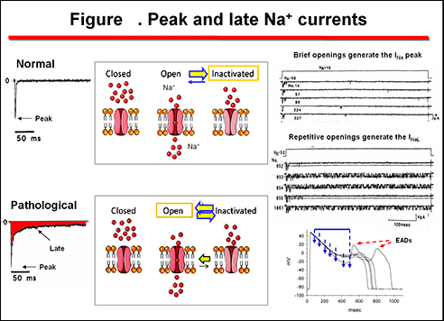
Figure shows that under normal conditions almost 99% of cardiac Na+ channels open transiently (1-3 ms), but rapidly inactivate-close and remain closed during the plateau phase of the AP. This short opening genetrates the peak Na+ currents (INa) that is responsible for the phase 0 of the cardiac AP and the QRS complex of the ECG. However, some Na+ channels present a slow or incomplete inactivation, so that that the inactivated channels can move from the open to the inactivated state and from the inactivated to the open state. As a consequence, the gating of the channel moves from isolated brief openings that generate the peak INa, to repetitive (bursts) of openings that generate the late Na+ current (INa,L). The amplitude of the INa,L is ~1% of the peak INa , but because it persists hundreds of milliseconds, the amount of Na+ carried by the INa,L can be of the same order as that carried by the peak INa (Zaza et al., 2008).

The INaL generated through open channels at the plateau level (i.e., phase 2) can be markedly increased under pathological conditions (i.e, ischemia, heart failure, LQTS3), leading to a prolongation of the ventricular APD (QT interval of the ECG) (Zaza et al., 2008). Mutations in the linker between domains III and IV in SCN5A, linked to the LQTS3, disrupt Na+ channel inactivation (Bennett et al., 1995). These “gain of function” mutations increase the amplitude of INaL, prolong the APD and may allow for the development of arrhythmogenic triggered activity, refered to as early afterdepolarizations (EADs). The enhanced inward current can be measured during sustained depolarizations and appears to reflect a change in channel gating that result in channel “bursting” (Bennett et al., 1995).

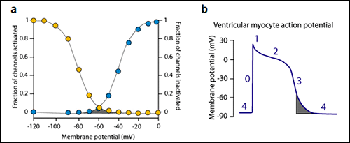
Additionally, a very small fraction of Na+ channels may reactivate during the phase 3 of the action potential. The probability of Na+ channel opening during the phase 3 of the cardiac AP is determined by the crossover of the activation and inactivation curves which govern the opening of the sodium channel (Figure xa) (Attwell et al., 1979). At the membrane potentials at which this crossover occurs a fraction of channels have recovered from inactivation and may reopen generating a new propagated response at the end of phase 3 of repolarization (Figure xb). The contribution of this window current under physiological circumstances is very limited because the overlap is less than 5% of the maximum current in healthy myocytes. However, under pathological conditions, some mutations of the Na+ channels can shift of voltage dependence of steady-state inactivation toward more depolarized membrane potentials leading to an increased “window” inward sodium current that can can lead to early afterdepolarizations arising at the end of the phase 3 of the cardiac AP (Amin et al., 2010).

Figure . The window sodium current. a) The window current (gray area) is determined by the crossover of the activation and inactivation curves which govern the opening of the sodium channel. B) This crossover takes place during the late phase 3 of repolzarization.

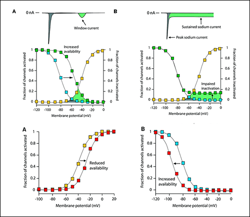
Figure. Effects of sodium channel mutations on channel gating.
Upper panels. A) A shift of the voltage dependence of inactivation curve to more depolarized membrane potentials leads to an increased “window” of inward sodium current. B) Tthe failure of the channel to inactivate completely lead to an increase in the late sodium current (INaL). Lower panels. Mechanism to reduce the peak sodium current. A: Some mutant channels shift of voltage dependence of steady-state activation toward more depolarized membrane potentials (from yellow to red) leading to a decrease in the INa. B) A hyperpolarizing shift of the voltage dependence of the steady-state inactivation curve to the left (from blue to red) also decreases the INa because, for a given resting membrane potential, fewer sodium channels are available.

Web de Interes Sanitario
Aviso legal

Esta obra está bajo una licencia de Creative Commons Reconocimiento-NoComercial-SinObraDerivada 4.0 Internacional
Licencia de Creative Commons